
Single-cell analysis of menstrual endometrial tissues defines phenotypes associated with endometriosis
Shih AJ, Adelson RP, Vashistha H, et al.BMC Medicine · 2022DOI 10.1186/s12916-022-02500-3
In plain language
Single-cell RNA sequencing of fresh menstrual effluent, collected at home with a menstrual cup, from 33 women: 11 with surgically confirmed endometriosis, 13 with symptoms but no diagnosis, and 9 controls. Uterine natural killer cells made up about 8% of cells in women with endometriosis against about 28% in controls, a decidualized stromal cell subset was less abundant in endometriosis, and the symptomatic group showed similar immune shifts.
Key numbers
- 33
- Women: 11 with endometriosis, 13 with symptoms, 9 controls
- 8% vs 28%
- Approximate share of uterine NK cells, endometriosis versus controls
- 43,054
- Cells analyzed after quality control
What it means
Shed endometrial tissue in menstrual effluent carries cell-level differences linked to endometriosis, including fewer uterine NK cells and fewer decidualized stromal cells. The authors note that single-cell sequencing is too costly to become a test itself, but it points to markers a simpler test could use.
What it does not show
A small study of fresh, live samples from a menstrual cup, shipped cold overnight, not dried samples. The symptomatic group had no surgical diagnosis, so the study could not measure how well these profiles predict endometriosis.
Figures
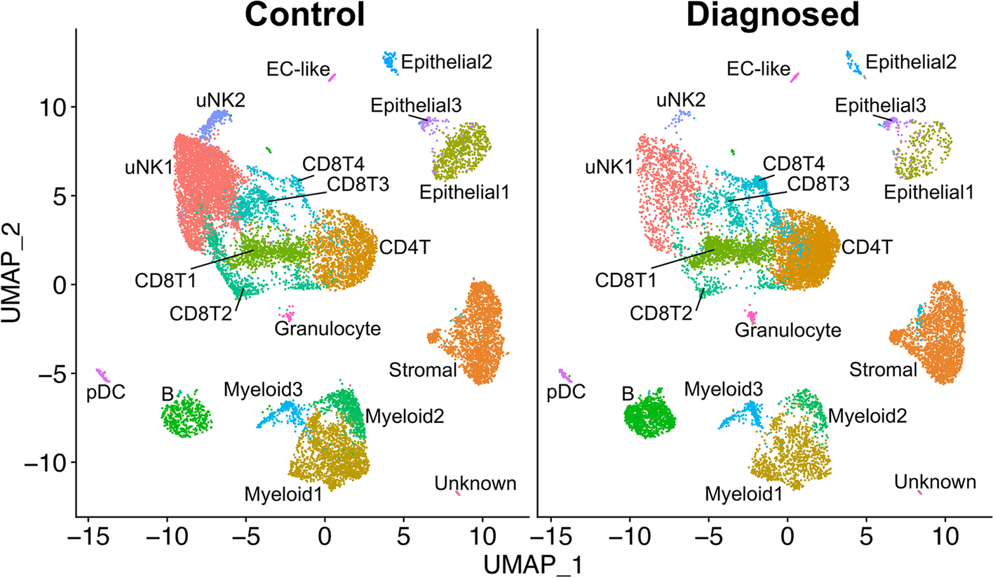
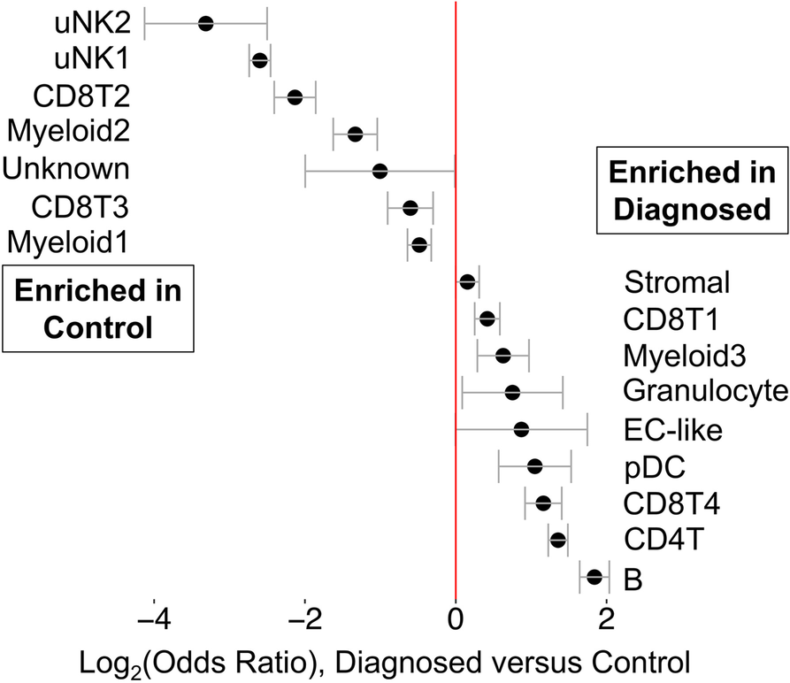
Figures reproduced unchanged from Shih AJ et al., BMC Medicine, 2022, under CC BY 4.0. Captions written by Qvin.
How to cite
Shih AJ, Adelson RP, Vashistha H, Khalili H, Nayyar A, Puran R, et al. Single-cell analysis of menstrual endometrial tissues defines phenotypes associated with endometriosis. BMC Medicine. 2022;20(1):315. doi:10.1186/s12916-022-02500-3